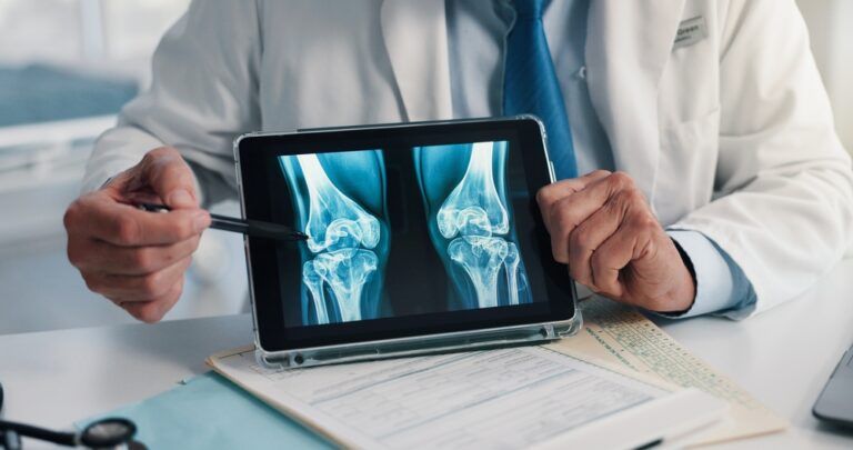
Exploring the Advancements in Digital X-Ray Technology

Digital X-ray technology has revolutionized modern dentistry, offering faster, safer, and more detailed imaging than traditional radiography. At Sunset Dental Group in Santa Ana, CA, Drs. Amanda and Jerry Kronquist emphasize the importance of patient education, and understanding how digital X-rays work can empower individuals to make informed decisions about their oral health. From diagnosis to treatment planning, the precision of digital imaging plays a crucial role in enhancing patient care.
As dental practices transition away from film-based systems, digital radiography is quickly becoming the gold standard. But beyond the convenience and speed, what truly sets digital X-rays apart are the advanced features and diagnostic capabilities they bring to the dental chair. This blog will walk through how digital X-rays work, the different types, their advantages, and how they support comprehensive dental care.
How Digital X-Rays Work
Unlike traditional film X-rays, digital radiography captures images using electronic sensors instead of photographic film. These sensors are placed inside the mouth and transmit the image to a computer screen almost instantly. The digital format allows for real-time viewing and manipulation, such as adjusting brightness or contrast to better detect abnormalities.
The process begins with positioning a sensor where the image is needed—typically around a tooth or jaw area. Once activated, the sensor captures X-ray photons and converts them into digital signals. These signals are processed and displayed on a monitor, allowing for immediate evaluation. Because the images are digitized, dentists can zoom in, highlight areas of concern, and even share the visuals with patients during the same appointment.
The shift from film to digital imaging represents a significant leap not only in convenience but also in clinical accuracy. By reducing the margin for human error and eliminating the need for chemical film development, digital radiography offers a cleaner, faster, and more precise method for capturing diagnostic images.
Types of Digital Dental X-Rays
Digital dental X-rays come in several forms, each designed to provide a different view or level of detail depending on the diagnostic need. Understanding the differences can help patients appreciate the value of each technique and reduce anxiety about the procedure.
Intraoral X-Rays
These are the most common and include:
- Bitewing X-rays: Capture the upper and lower teeth in one area of the mouth. They are ideal for detecting decay between teeth and monitoring bone levels.
- Periapical X-rays: Focus on one or two teeth at a time, showing the entire tooth from crown to root. These are especially useful for identifying root infections or abscesses.
- Occlusal X-rays: Offer a broader view of the floor or roof of the mouth and are often used in pediatric dentistry.
Extraoral X-Rays
These are taken outside the mouth and include:
- Panoramic X-rays: Capture a full view of the jaw, sinuses, and temporomandibular joints. Ideal for evaluating wisdom teeth, bone abnormalities, or planning implants.
- Cephalometric X-rays: Often used in orthodontics to assess jaw alignment and plan braces.
Each type serves a unique purpose and may be used in combination to form a comprehensive view of a patient’s oral health.
Benefits Over Traditional Radiography
One of the most significant advantages of digital X-ray technology is the reduced exposure to radiation. Studies show that digital systems can reduce radiation by up to 80% compared to film-based systems. This is particularly beneficial for children, pregnant women, or patients who require frequent imaging.
Another key benefit is image quality. Digital X-rays offer high-resolution images that can be enlarged, enhanced, and stored electronically. This makes it easier to identify cavities, bone loss, or infections at an earlier stage. Since these images are instantly available, they also streamline the workflow in a dental office, cutting down on appointment times and allowing for faster diagnosis and treatment planning.
Additionally, digital radiographs are environmentally friendly. They eliminate the need for chemical developers and film, reducing hazardous waste and lowering the environmental footprint of dental practices.
Role in Diagnostic Accuracy and Treatment Planning
Digital X-ray technology significantly enhances diagnostic accuracy, giving dentists clearer insight into oral structures and potential issues. With the ability to manipulate image properties, practitioners can reveal details that would be difficult to detect using traditional film.
For instance, digital images allow for contrast adjustments that can highlight early signs of tooth decay or periodontal disease. The high-definition visuals also help with identifying cracks, root fractures, or impacted teeth. This leads to earlier and more accurate diagnoses, improving patient outcomes and reducing the need for invasive procedures.
Digital X-rays also support comprehensive treatment planning. Dentists can use the images to map out surgical procedures, monitor healing progress, and track changes over time. In complex cases such as dental implants, orthodontics, or full-mouth restorations, the accuracy and detail provided by digital imaging are essential.
Integration with Modern Dental Technology
Another advantage of digital X-rays is their compatibility with other digital tools used in modern dentistry. Many dental software programs integrate seamlessly with digital imaging systems, allowing for better case documentation and interdisciplinary communication.
For example, 3D imaging technologies like cone-beam computed tomography (CBCT) build upon digital radiography to provide volumetric data. These tools are often used for implant planning, endodontic diagnosis, or assessing TMJ disorders. Digital X-rays can also be used in conjunction with CAD/CAM systems, which are used to design and fabricate crowns, bridges, and veneers.
This integration streamlines the dental workflow, enhances precision, and promotes a more collaborative approach to patient care. For patients, this means fewer appointments, better treatment outcomes, and a clearer understanding of their oral health.
What to Expect During a Digital X-Ray Appointment
For many patients, one of the most reassuring aspects of digital X-rays is their speed and comfort. The sensors used are smaller and more comfortable than traditional film, and the process is usually completed within minutes.
Here’s what typically happens during a digital X-ray visit:
- The dental assistant places a lead apron over your chest and neck for protection.
- A small digital sensor is gently placed inside the mouth.
- You are asked to bite down or hold still while the X-ray is taken.
- The image appears instantly on a screen for review.
Patients are generally surprised by how quick and noninvasive the process is. There’s no waiting for film to develop, and the entire appointment moves along more efficiently thanks to the immediate image availability.
Final Thoughts on the Future of Dental Imaging
As technology continues to evolve, digital X-rays will likely become even more advanced, incorporating artificial intelligence and machine learning to aid in diagnosis. This future promises faster detection of anomalies and greater consistency in interpreting results.
At Sunset Dental Group in Santa Ana, CA, Drs. Amanda and Jerry Kronquist encourage patients to stay informed about the technologies used in modern dentistry. While digital X-rays are just one part of comprehensive care, they play a central role in ensuring accurate diagnoses and well-informed treatment decisions. Understanding how these tools work can make dental visits more transparent and less intimidating for patients of all ages.
Resources
Jacobs, R., Salmon, B., Codari, M., Hassan, B., & Bornstein, M. M. (2018). Cone Beam Computed Tomography in Implant Dentistry: Recommendations for Clinical Use. BMC Oral Health.
Wenzel, A. (2006). Digital Radiography and Caries Diagnosis. Dentomaxillofacial Radiology.
Farman, A. G., & Farman, T. T. (2005). A Comparison of 18 Different X-Ray Detectors Currently Used in Dentistry. Oral Surgery, Oral Medicine, Oral Pathology, Oral Radiology, and Endodontology.

Recent Comments